MicroCT imaging of the zebrafish – a surprisingly good genetic model of human diseases
The zebrafish (Danio rerio) preclinical-genetic model of chordate phenotype and disease is not new. Scientists have been using it for biomedical research for decades, taking advantage of its short lifecycle and rapid rate of reproduction to allow selection and multiplication of genotypes much faster than the mouse1.
Kwon, Watson and Karasik in their 2019 paper “Using zebrafish to study skeletal genomics” summed up the reason that the zebrafish is so useful: “Due to their amenability to rapid genetic approaches, as well as the large number of conserved genetic and phenotypic features, there is a strong rationale supporting the use of zebrafish for human skeletal genomic studies2.”
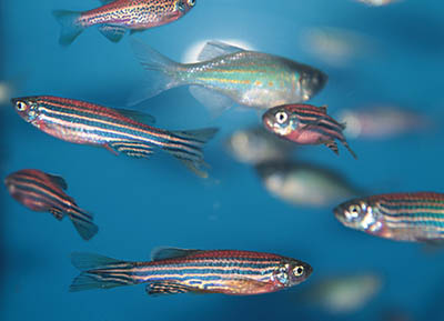 Figure 1. The zebrafish (Danio rerio) is a well-established preclinical model useful in genetics due to its fast generation time. Zebrafish are particularly well suited for rapid generation of mutant lines for selected genes, and a rapidly increasing number of human disease models are being successfully made with the zebrafish. This began with bone diseases but is extending to many soft tissue diseases2. Due to their small size the zebrafish are also convenient to scan by microCT. For bone imaging no special preparation is needed while imaging of soft tissue is facilitated by the usual soft tissue contrast agents such as phosphotungstic or phosphomolybdenic acid, osmium tetroxide and iodine (Figure 2). 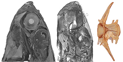 Figure 2. The zebrafish head scanned by microCT (SKYSCAN 2214) after phosphoungstic acid (in ethanol) staining (left) and without staining (center); the zebrafish vertebra (right). Arthritis can be accurately modelled by a zebrafish strain which shows vertebral degeneration more similar to human arthritis than mouse models (Figure 3). And among the soft tissue diseases that can be modelled by the zebrafish is tuberculosis (Figure 4). 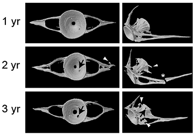 Figure 3. A zebrafish model of osteoarthritis featuring vertebral degeneration3. 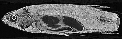 Figure 4. Zebrafish model of tuberculosis; the white particles around the gut lining are tuberculosis nodules. Courtesy of Anna Bruinen and Ron Heeren, IMS group (M41), University of Maastricht. Bruker microCT scanners such as the SKYSCAN 1272 and 1275 are ideally suited to high throughput imaging of zebrafish ex vivo, with their desktop ease-of-use and large camera field of view allowing whole fish to be scanned rapidly at good resolution. This is further facilitated by the robotic auto-sample changer on both these scanners. For the ultimate resolution, zebrafish can be imaged in the SKYSCAN 2214 nanoCT with stable and repeatable results below the micron level.
How can a fish usefully model human diseases? In the context of the whole diverse animal kingdom, fish and humans are relatively close relatives. It turns out that the genetics of many diseases is remarkably conserved over evolutionary history. And consider the hormone estrogen – central to the control of bone metabolism: it evolved in the very first chordates at the “Cambrian explosion” of multicellular life, 500 million years ago4. More and more preclinical disease models are thus being realised in the zebrafish2.
Read MN127_Zebrafish exvivo microCT imaging and analysis to learn more.
References
- Streisinger G et al. (1981) Production of clones of homozygous diploid zebra fish (Brachydanio rerio). Nature 291 (5813) 293–296.
- Kwon R et al. (2019) Using Zebrafish to Study Skeletal Genomics. Bone 126: 37-50.
- Hayes AJ et al. (2013) Spinal deformity in aged zebrafish is accompanied by degenerative changes to their vertebrae that resemble osteoarthritis. PloS one. 8(9): e75787.
- Callard GV et al. (2011) Evolutionary origins of the estrogen signaling system: insights from amphioxus. J Steroid Biochem Mol Biol. 127(3-5): 176–188.
|
