The new NRecon version 2.0. features phase retrieval imaging
Our July newsletter is close in time to the release of a major new version of our reconstruction software NRecon – version 2.0. This has many important new capabilities and enhancements, and one of these is phase retrieval 3D reconstruction. The phase retrieval method of Paganin (1) is one of a class of techniques termed “phase contrast”. X-ray phase contrast imaging makes use of the phenomena of refraction and diffraction of X-rays at internal boundaries of a sample, and is thus complementary to conventional absorption contrast. This is equivalent to the phase contrast imaging that is performed with visible light microscopes. Phase interactions of X-rays are of smaller magnitude than those that occur with visible light, but they are significant enough to provide the basis for realising X-ray phase contrast.
Paganin phase retrieval imaging
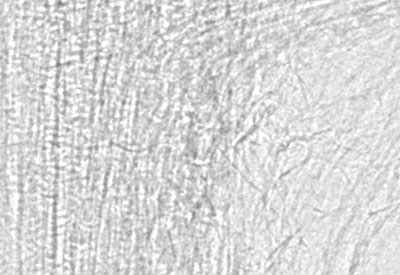
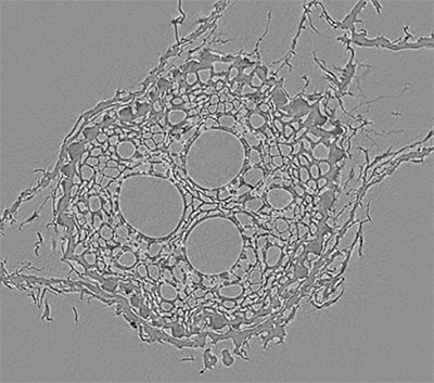
Some phase contrast methods require synchrotron radiation since they need X-rays to be of a single energy only (monochromatic) and coherent (“in step”, in the same wavetrain phase). Phase contrast methods sometimes employ precise gratings, or alternatively multiple scans of different source-camera distance, to generate the needed phase effects. This all adds to the complexity of phase contrast X-ray imaging. However, there is one phase contrast phenomenon that does happen readily in laboratory microCT systems even with polychromatic and noncoherent laboratory sources, and without the need of any gratings; this is small angle scattering. This phenomenon results in a bright “halo” surrounding object boundaries in both the projection and the reconstructed images as shown in a submicron scan of a Sorhgum plant root in figure 1. It results from the reflection of some X-rays that are incident at an object surface at a very shallow angle, much smaller than one degree.
(a) (b) (c)
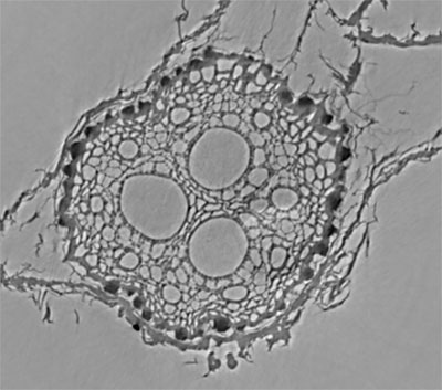
Figure 1. From the scan of reconstructed cross-sections of a root of Sorghum bicolor, a part of a projection image (a) and a standard reconstructed cross-section (b) show the bright edges characteristic of small angle scattering. Such bright edges indicate the potential for phase retrieval reconstruction (PR). The PR reconstruction of the same scan (c) show the conversion of the edge signal into enhanced contrast, revealing for example the higher density of a layer of peripheral cells in the root (arrow), which is not visible in the reconstruction of standard absorption based projections. Courtesy of Dr Rivka Elbaum, Faculty of Agriculture, Hebrew University of Jerusalem, Israel.
Phase retrieval (Paganin) is a method that obtains enhanced sample contrast and structural information from this small angle scattered component, and it can be realized in a single scan, not requiring multiple geometries or gratings (1). Phase retrieval is thus applicable to scans at high resolution in laboratory (including desktop) microCT scanners, and this capability has now been added to Bruker NRecon. Please note that phase retrieval is not a different CT reconstruction algorithm. It is an image processing method applied to individual 2D X-ray projection (and other) images. By applying it to a full set of scan projections, micro and nanoCT can be performed with phase retrieval.
The results of this phase retrieval applied to the sorghum root scan is also shown in figure 1 (c). The notable feature of phase retrieval imaging is the elimination of the artificial edge brightening and its conversion into enhanced contrast. Thus for example the higher density of a layer of peripheral cells in the root is shown by phase retrieval, but is not visible in the reconstruction of standard absorption based projections
How to perform phase retrieval in NRecon 2.0
How is phase retrieval done? Some users may be familiar with an ImageJ plugin called “Ankaphase” which performs Paganin phase retrieval on images including CT scan projections. NRecon version 2 has a similar new function which allows you to convert the full set of a scan’s projection images to their phase-retrieved counterparts. However it’s fair to say that the NRecon dialog window for doing this – shown below in figure 2 – is quite a lot simpler than ImageJ’s Ankaphase since many parameters are silently read from the scan log file. The critical parameter that you have to enter is the “delta/beta ratio” which relates to the strength of phase retrieval. One typically enters values from 100-1000, with 400-700 often being a good starting point. Other settings can – at first at least – be left at default values.
So it’s quite easy to get started with phase retrieval imaging of your own scans. High resolution scans with noticeable edge brightening are most likely to give useful results with phase retrieval; if in doubt, just try it. And since nothing additional is needed, even existing scans you have already done can be reconstructed with phase retrieval if you still have the scan projection images.
Full details of the phase retrieval method including an overview of its theory and how to do it step by step in NRecon 2.0 are given in the new method note MN133 that is linked with this newsletter, entitled: “MN 133 Paganin single-distance phase retrieval reconstruction in NRecon”.
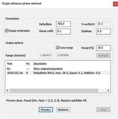
Figure 2. This is the new window in NRecon 2.0 for making phase retrieval transformation of projection datasets. The main parameter to adjust is the delta/beta ratio. You process your scan’s projections using this window, with single projection preview first. The processed projections are written to a subfolder named “PR”; load and reconstruct PR cross-sections from these phase-retrieved projections, in NRecon. References
- Paganin D, Mayo SC, Gureyev TE, Miller PR, Wilkins SW (2002) Simultaneous phase and amplitude extraction from a single defocused image of a homogeneous object. J. Microsc. 206, 33-40.
- The company collaboration between the company RJL Micro & Analytical and Helios, in German language on RJL’s website.
|
