In this issue
|
|
▪ The morphological escalator
▪ Image of the month
▪ Bruker microCT news
▪ Upcoming events
|
The morphological escalator – a new technique for automatic trabecular-cortical separation Bone morphometry in the mouse and rat preclinical models requires separation of the medullary trabecular bone from surrounding cortical bone at the endosteal boundary. This can be done by manual drawing of region of interest (ROI) shapes. However advanced image processing methods also allow this trabecular delineation to be automated. A method note already exists, MN008, entitled “Automated trabecular and cortical bone selection”, which shows how morphological (erode, dilate, open, close), despeckle and bitwise (Boolean) image operations can be combined to effectively separate trabecular from cortical bone at the rodent metaphyseal medulla.
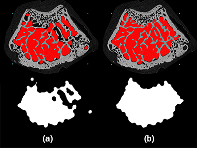
Here we introduce a modified version of this method using a technique called the “morphological escalator”. This addresses a difficult challenge to automatic trabecular delineation where parts of the cortical wall are highly porous with only thin structures. Such regions include peripheral growth plate remnants that persist some distance downstream of the growth plate, especially in young fast-growing rodents. Such regions are shown in figure 1 at three locations in a mouse femur metaphysis cross-section.
Trabecular bone auto-separation methods are based on morphological operations: erode (remove a pixel layer from surface), dilate (add a pixel layer to surface), open (erode then dilate) and close (dilate then erode). The medullary space can be binarised, and gaps from trabecular structures closed out, to make a trabecular volume of interest (VOI). What complicates this, however, is overlap in thickness between trabecular and cortical bone. This means that a single combination of opening and closing sometimes does not cleanly separate trabecular bone, but creates artefacts.
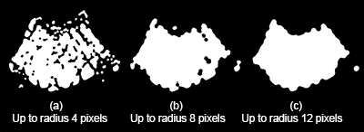
A solution to this is the “morphological escalator”. This is a series of open-close operations, starting with a voxel radius of 1, then repeating with a successively increasing voxel radius up to a value depending on the thickness of the trabecular structures (open 1 pixel; close 1 pixel; open 2 pixels; close 2 pixels, etc.). In figure 1 the effects of opening and closing are shown to a radius of 12 voxels, comparing a single open-close morphological operation (a) to the stepwise morphological escalator (b).
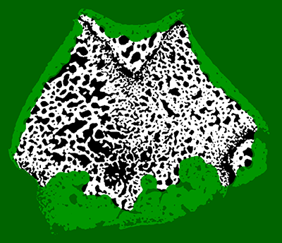
The morphological escalator appears in figure 1 to do quite a good job of excluding from the trabecular VOI peripheral remnants of growth plate – places in the cortical boundary which are fine textured and highly porous. This restricts the analysis to mature secondary trabecular bone. Other trabecular-cortical separation algorithms, while otherwise effective, do not always succeed in excluding these peripheral growth plate remnants. Figure 2 illustrates how the morphological escalator works. Regions become progressively solidified as white or black depending on the local percentage volume and relative thickness of bone or soft tissue spaces.
|
|
|
|
|
|
Figure 1. Delineation of a trabecular VOI involves morphological operations such as opening and closing. A single morphological operation with a high radius, however, causes unwanted gaps, as seen in the images to the left (a). The “morphological escalator” – a series of opening and closing operations with a stepwise increasing voxel radius – achieves a better, more continuous result (b).
|
|
|
|
Figure 2. The morphological escalator progressively solidifies regions of medulla and cortical wall. Some isolated shapes are created by the end, but these can be removed by despeckling (an object labeling operation).
|
|
|
|
The method 124 Trab-cort bone auto-separation with the morphological escalator shown here using the morphological escalator is useful for mouse bone with relatively sparse trabeculae, and excludes peripheral growth plate remnants. However it does not work as well with highly dense medullary regions of rat bones where percent trabecular volume can be above 50% (see image above). In this case the exclusion of peripheral growth plate remnants is less effective. Other approaches, such as manual delineation, would be needed in such cases.
|
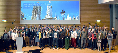
Image of the Month Bruker microCT 2019 User Meeting |
|
|
|
|
|
|
|
Volume 6, Issue 3
June 2019
|
Bruker microCT news
|
| - microCT User Meeting
Another year, another user meeting. This year’s gathering in the medieval town of Mechelen, Belgium was a feast of history and architecture, world class science and beer tasting at a historic Belgian brewery.
Eighty participants from 25 countries wove a web of networking while sharing cutting edge developments in 3D imaging, including oil-water flow analysis, neural network screening for shape, deep learning of textural clues to invisible features, phase retrieval, new cartilage analysis methods, and more.
As usual we had our competitions – Anna Jacobsen from California won best poster, while Liebert Parreiras Nogueira from Oslo won best microCT movie.
Visit our website for more information!
- BrukerSupport: Software Updates & Method Notes
We have pre-registered our users at BrukerSupport.com for the distribution of documentation and software. Please log into your account and select your SKYSCAN microCT systems.
At BrukerSupport.com you'll find the latest software downloads available for you. In addition, the complete library of method notes has been uploaded as well.
If you did not receive an email for log-in, please apply for an account with your system serial number. Enjoy your support membership and let us know if you have any questions!
|
|
Upcoming events
|
| Bruker microCT will participate with an exhibit in these upcoming conferences. Please click the links below for more information. We hope to see you there! |
| June 19 - 22, 2019
IADR/AADR/CADR - International Association for Dental Research - Vancouver, Canada June 25 - 26, 2019
dXCT - Dimensional X-ray Computed Tomography - Huddersfield, UK July 02 - 04, 2019
DIR – Symposium on Digital Industrial Radiology and Computed Tomography – Fürth, Germany July 22 - 24, 2019
URTeC – Unconventional Resources Technology Conference – Denver, CO, US July 22 - 26, 2019
ICTMS - International Conference on Tomography of Materials & Structures - Cairns, Australia August 05 - 09, 2019
DXC - Denver X-Ray Conference - Chicago, IL, US September 04 - 07, 2019
WMIC – The World Molecular Imaging Congress – Montréal, QC, Canada September 20 - 23, 2019
ASBMR – The American Society for Bone and Mineral Research – Orlando, FL, US |
|
|
|